Science, human bodyLabelled diagramPart 1 of 8, The Human HeartUpdated 1 October 2026
The Human Heart Explained
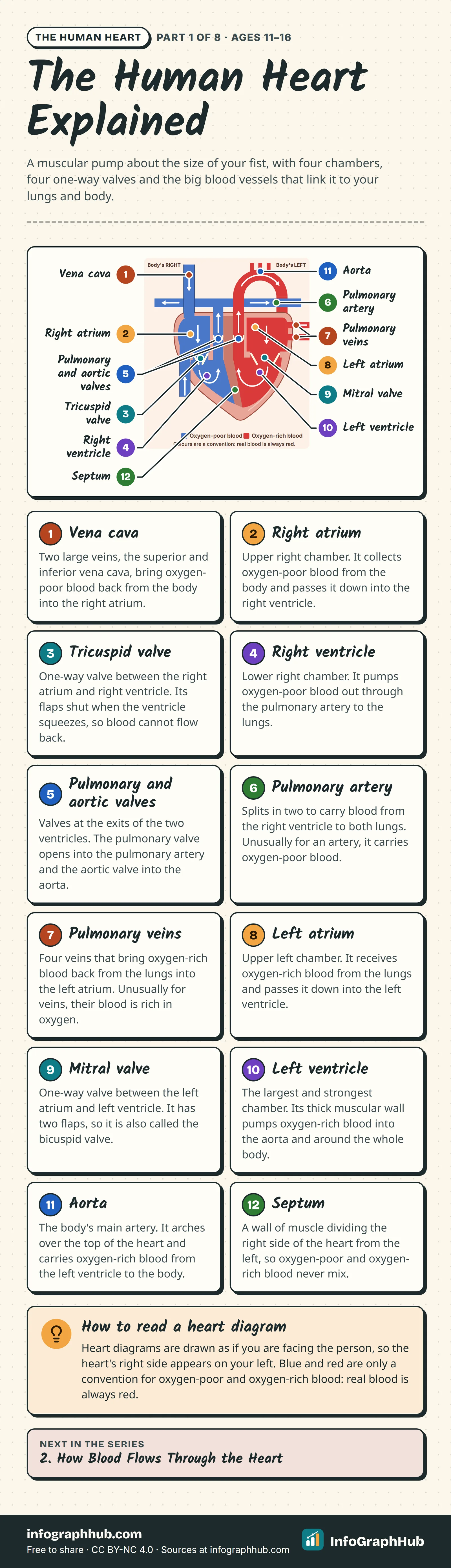
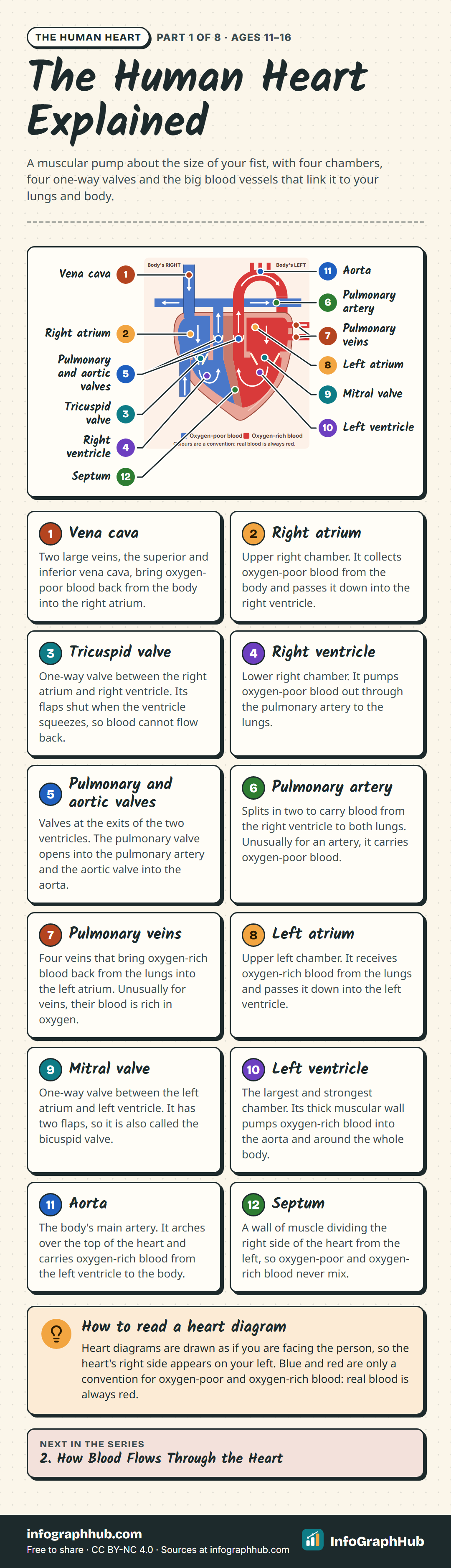
The heart is a muscular pump in the middle of your chest. A labelled front-view diagram of its four chambers, four one-way valves, the septum and the great blood vessels that link it to the lungs and the rest of the body.
Interactive
Explore it step by step
12 labelled parts: Vena cava → Right atrium → Tricuspid valve → Right ventricle → Pulmonary and aortic valves → Pulmonary artery → Pulmonary veins → Left atrium → Mitral valve → Left ventricle → Aorta → Septum. Press play, or choose a number to explore.
Step 1 of 12: Vena cava
Two large veins, the superior and inferior vena cava, bring oxygen-poor blood back from the body into the right atrium.
Step 2 of 12: Right atrium
Upper right chamber. It collects oxygen-poor blood from the body and passes it down into the right ventricle.
Step 3 of 12: Tricuspid valve
One-way valve between the right atrium and right ventricle. Its flaps shut when the ventricle squeezes, so blood cannot flow back.
Step 4 of 12: Right ventricle
Lower right chamber. It pumps oxygen-poor blood out through the pulmonary artery to the lungs.
Step 5 of 12: Pulmonary and aortic valves
Valves at the exits of the two ventricles. The pulmonary valve opens into the pulmonary artery and the aortic valve into the aorta.
Step 6 of 12: Pulmonary artery
Splits in two to carry blood from the right ventricle to both lungs. Unusually for an artery, it carries oxygen-poor blood.
Step 7 of 12: Pulmonary veins
Four veins that bring oxygen-rich blood back from the lungs into the left atrium. Unusually for veins, their blood is rich in oxygen.
Step 8 of 12: Left atrium
Upper left chamber. It receives oxygen-rich blood from the lungs and passes it down into the left ventricle.
Step 9 of 12: Mitral valve
One-way valve between the left atrium and left ventricle. It has two flaps, so it is also called the bicuspid valve.
Step 10 of 12: Left ventricle
The largest and strongest chamber. Its thick muscular wall pumps oxygen-rich blood into the aorta and around the whole body.
Step 11 of 12: Aorta
The body's main artery. It arches over the top of the heart and carries oxygen-rich blood from the left ventricle to the body.
Step 12 of 12: Septum
A wall of muscle dividing the right side of the heart from the left, so oxygen-poor and oxygen-rich blood never mix.
Use this infographic
Free under CC BY-NC 4.0, with credit to InfoGraphHub.
What the heart is and where it sits
The heart is a hollow organ made mostly of muscle. Its job is to pump blood through a network of blood vessels to every part of the body, without stopping, for your whole life.
It sits in the middle of your chest, between the two lungs and behind the breastbone, tilted slightly to the left. Its rounded top, where the big blood vessels join it, is called the base. Its narrow lower tip, the apex, points down and to the left. That tilt is why most people feel their heartbeat a little to the left of centre.
Two pumps side by side
The heart is really two pumps joined together. A wall of muscle called the septum divides it into a right side and a left side, so the blood in the two halves never mixes.
- The right side receives oxygen-poor blood from the body and pumps it to the lungs, where it picks up oxygen.
- The left side receives oxygen-rich blood from the lungs and pumps it to the rest of the body.
Both sides beat at the same moment, so every heartbeat drives both journeys at once. The step-by-step route is the subject of the next part of this series.
Four chambers
Each side has two hollow chambers:
- An atrium (plural atria) at the top. The atria are receiving chambers: blood flows into them from the veins.
- A ventricle at the bottom. The ventricles are the main pumping chambers: they squeeze blood out into the arteries.
The two ventricles push out the same amount of blood with each beat, but the left ventricle has a much thicker muscle wall. It has to build up far more pressure, because it pushes blood around the whole body, while the right ventricle only has to reach the nearby lungs.
Four one-way valves
Valves are flaps of tough tissue that let blood through in one direction only. Each one opens when the pressure behind it is higher and snaps shut if blood starts to flow backwards.
| Valve | Where it is |
|---|---|
| Tricuspid | between the right atrium and right ventricle |
| Pulmonary | between the right ventricle and the pulmonary artery |
| Mitral (bicuspid) | between the left atrium and left ventricle |
| Aortic | between the left ventricle and the aorta |
The tricuspid and mitral valves are held in place by strong cords called chordae tendineae, which stop the flaps from being pushed back up into the atria when the ventricles squeeze.
The great blood vessels
Four kinds of large blood vessel connect the heart to the body and lungs:
- Vena cava: the superior (upper) and inferior (lower) vena cava bring oxygen-poor blood from the body into the right atrium.
- Pulmonary artery: carries oxygen-poor blood from the right ventricle to the lungs.
- Pulmonary veins: four veins that bring oxygen-rich blood from the lungs into the left atrium.
- Aorta: the body's main artery, which carries oxygen-rich blood from the left ventricle to the rest of the body.
A simple rule helps: arteries carry blood away from the heart and veins bring it back.
The heart's walls and its own blood supply
The heart wall has three layers: a thin inner lining (endocardium), a thick middle layer of heart muscle (myocardium) that does the squeezing, and an outer protective sac (pericardium) with a little fluid in it to reduce friction as the heart moves.
Heart muscle works non-stop, so it needs its own supply of oxygen. The blood inside the chambers does not feed it. Instead, the coronary arteries branch off the aorta and spread over the outside of the heart, delivering oxygen-rich blood to the muscle itself.
Reading a heart diagram
Heart diagrams are drawn as if you are facing the person, so the heart's right side appears on your left. Always work out which side is which before you label anything.
Diagrams also colour oxygen-poor blood blue and oxygen-rich blood red. This is only a convention to tell the two apart: real blood is always red, darker when it carries less oxygen.
Frequently asked questions
Where exactly is your heart?
In the middle of your chest, between your lungs and behind your breastbone, tilted slightly to the left. Its lower tip points down and to the left, which is why you feel the beat just left of centre.
Why is the wall of the left ventricle thicker than the right?
Both ventricles pump the same amount of blood with each beat, but the left ventricle has to push it all the way around the body. That needs much more pressure than the short trip from the right ventricle to the lungs, so its muscle is thicker.
Why do heart diagrams look back to front?
They show the heart as if you are facing the person. The heart's right side is therefore on the left of the page, and its left side is on the right.
What do the four heart valves do?
They keep blood moving in one direction. The tricuspid and mitral valves stop blood flowing back from the ventricles into the atria, and the pulmonary and aortic valves stop it flowing back from the arteries into the ventricles.
Why are diagrams of the heart coloured red and blue?
It is a convention: blue marks oxygen-poor blood and red marks oxygen-rich blood, so you can follow the two streams. Real blood is red in both cases, just darker when it carries less oxygen.

Sources & methodology
Every fact is checked against the sources below. We write original explanations and draw original graphics; no figures are copied from textbooks. Spotted an error? See our corrections policy.
- How the Heart Works: Heart Anatomy (US National Heart, Lung, and Blood Institute (NHLBI), accessed 1 Oct 2026)
- How the Heart Works: How Blood Flows through the Heart (US National Heart, Lung, and Blood Institute (NHLBI), accessed 1 Oct 2026)
- How your heart works (British Heart Foundation, accessed 1 Oct 2026)
- Heart Anatomy (Texas Heart Institute, accessed 1 Oct 2026)
- Anatomy and Physiology 2e, 19.1 Heart Anatomy (OpenStax, Rice University, accessed 1 Oct 2026)
Editorial review pending · Updated
Made by the InfoGraphHub editorial team (how we research and check: editorial and AI policy). First published 1 October 2026, last reviewed 1 October 2026. Found a mistake? Tell the editors.
For teachers: ages and curriculum fit
Ages 11–16 · Grades 6–10 (US) · Years 7–11 (UK) · Classes 6–10 (India)
NGSS is a registered trademark of WestEd. Cambridge and IGCSE are trademarks of Cambridge University Press & Assessment. Names and codes are used only to describe curriculum fit; InfoGraphHub is not endorsed by these organisations.
Embed this on your site
Paste this HTML into your page. Keep the credit line: it is required by the CC BY-NC 4.0 license.
Related infographics


For teachers and parents: new InfoGraphHub resources by email
One short email when new free resources go live. For adults (18+) only. No spam, no tracking in emails, unsubscribe anytime.