8-part series · Ages 11–16
The Human Heart
Eight infographics on the pump that keeps you alive: its parts, the route of blood, the numbers, the history and the myths.
The 1 parts (7 more coming soon)
The complete guide
The Human Heart explained
Your heart has beaten around 100,000 times since this time yesterday. This series takes the heart one question at a time: what its parts are, which way the blood goes, how much it pumps, how arteries, veins and capillaries differ, which words to use, how scientists worked it all out, and which popular ideas are wrong.
Each part is a printable infographic with sources, written for ages 11–16 and their teachers. It is about how a healthy heart works, not medical advice. Work through the parts in order or jump to the one you need, then test yourself with the quiz at the end.
What the heart is and what it does
The heart is a hollow, muscular organ whose job is to pump blood around the body through a closed network of tubes, the blood vessels. Blood carries oxygen and food substances such as glucose to every cell and carries waste, including carbon dioxide, away. Because cells need a constant supply, the heart has to keep working, beat after beat, for your whole life.
The heart sits in the middle of the chest, between the two lungs and behind the breastbone, tilted so that its lower tip, the apex, points down and to the left. An adult heart is about the size of a fist. Its wall has three layers: a thin inner lining (the endocardium), a thick middle layer of heart muscle (the myocardium) that does the pumping, and a protective outer sac (the pericardium).
Heart muscle needs oxygen too, and it does not take it from the blood passing through its chambers. Instead, the coronary arteries branch off the aorta and run over the surface of the heart to feed the muscle itself.
Four chambers, four valves and the septum
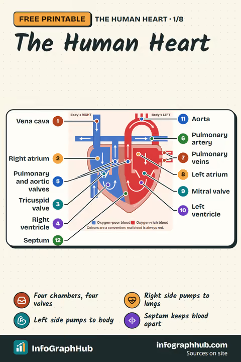
The heart is really two pumps side by side. A wall of muscle, the septum, divides it into a right side and a left side so that the blood in the two halves never mixes.
Each side has two chambers:
- an atrium at the top, which receives blood from the veins;
- a ventricle at the bottom, which pumps blood out into the arteries.
Four valves keep the blood moving one way:
| Valve | Position | Type |
|---|---|---|
| Tricuspid (3 flaps) | right atrium → right ventricle | atrioventricular |
| Pulmonary | right ventricle → pulmonary artery | semilunar |
| Mitral or bicuspid (2 flaps) | left atrium → left ventricle | atrioventricular |
| Aortic | left ventricle → aorta | semilunar |
Strong cords called chordae tendineae anchor the atrioventricular valves so the flaps cannot be pushed back into the atria.
When you label a heart, remember that diagrams show it as if you are facing the person: the heart's right side is on the left of the page. Diagrams also colour oxygen-poor blood blue and oxygen-rich blood red. That is only a convention; real blood is always red.
The route blood takes
Humans have a double circulation: two loops that meet at the heart.
- Oxygen-poor blood from the body enters the right atrium through the superior and inferior vena cava.
- It passes through the tricuspid valve into the right ventricle.
- The right ventricle pumps it through the pulmonary valve into the pulmonary artery, which takes it to the lungs.
- In the lungs it gives up carbon dioxide and picks up oxygen.
- Oxygen-rich blood returns through the pulmonary veins into the left atrium.
- It passes through the mitral valve into the left ventricle.
- The left ventricle pumps it through the aortic valve into the aorta and round the body, and it eventually returns to the vena cava.
Steps 1 to 3 and the return to the left side form the pulmonary circulation; the trip from the left ventricle round the body and back is the systemic circulation. On every complete circuit, blood passes through the heart twice.
The two sides work at the same time. Both atria contract together, then both ventricles. So a single beat sends blood to the lungs and to the body at once. A fish, by contrast, has a single circulation: its two-chambered heart pumps blood through the gills and then straight on round the body, so the blood is not pumped again after it picks up oxygen.
What keeps the beat going
Every heartbeat starts with an electrical signal from the sinoatrial (SA) node, a small group of pacemaker cells in the wall of the right atrium. The signal spreads across both atria, which contract and push blood down into the ventricles. It then reaches the atrioventricular (AV) node, which holds it back for a fraction of a second so the ventricles can finish filling. Finally the signal spreads through the walls of the ventricles, which contract and pump blood out.
The contracting phase of a beat is called systole and the relaxing, filling phase diastole. At 75 beats a minute, one full cycle takes about 0.8 seconds. Left completely alone, the SA node would fire roughly 80 to 100 times a minute; nerves from the brain and hormones such as adrenaline speed the heart up or slow it down to match what the body needs. The electrical activity can be recorded from the skin as an electrocardiogram (ECG), with waves labelled P, QRS and T.
The heart in numbers
A few figures give a sense of the heart's workload:
- Heart rate. A normal resting rate for adults and anyone aged 10 or over is 60 to 100 beats a minute. Younger children have faster rates, and very fit athletes can rest at 40 to 60.
- Beats per day. At about 70 beats a minute: 70 × 60 × 24 = 100,800, so roughly 100,000 beats a day.
- Stroke volume. Each ventricle pushes out about 70 mL of blood per beat at rest.
- Cardiac output. Stroke volume × heart rate gives the volume pumped per minute: 70 mL × 75 = 5,250 mL, or about 5 litres a minute. That is about 7,500 litres a day, although the body holds only about 5 litres of blood in total.
- Size. An adult heart is about 12 cm long and weighs roughly 250 to 350 grams.
During hard exercise, a healthy young person's heart rate can rise to about 150 beats a minute and the stroke volume to about 130 mL, raising the output to around 19.5 litres a minute: four to five times the resting figure.
Heart rate also changes with age. Resting ranges given by the US National Library of Medicine are:
| Age | Normal resting rate (beats per minute) |
|---|---|
| Newborn (up to 1 month) | 70–190 |
| 1–2 years | 80–130 |
| 5–6 years | 75–115 |
| 7–9 years | 70–110 |
| 10 years and over, adults | 60–100 |
These ranges describe healthy people at rest. Anyone's rate rises with exercise, excitement or nerves and falls during rest and sleep.
Arteries, veins and capillaries
Blood leaves the heart in arteries, which branch into ever-smaller vessels, then flows through capillaries and returns in veins. Each type of vessel is built for its job:
| Arteries | Capillaries | Veins | |
|---|---|---|---|
| Direction | away from the heart | link arteries to veins | back to the heart |
| Pressure | high | falling | low |
| Wall | thick, muscular and elastic | one cell thick | thinner, less muscle |
| Lumen | narrow | 5–10 micrometres | wide |
| Valves | none along their length | none | many, especially in the limbs |
The elastic walls of arteries stretch with each surge of blood and spring back, helping to keep it moving. Capillary walls are so thin that oxygen and glucose can pass out to the cells and carbon dioxide can pass in. Veins have valves because their blood is at low pressure and, in the legs, has to travel upwards against gravity.
The rule that defines an artery is direction, not oxygen. The pulmonary artery carries oxygen-poor blood to the lungs, and the pulmonary veins carry oxygen-rich blood back.
How we found out
Our picture of the heart took centuries to build:
- 2nd century AD. The Greek physician Galen taught that the liver made blood, that the body used it up, and that some blood seeped through invisible pores in the septum.
- 13th century. Working in Cairo, Ibn al-Nafis wrote that the septum has no pores and that blood from the right side travels through the lungs to the left: the earliest known description of the pulmonary circulation.
- 1628. William Harvey published De Motu Cordis. By multiplying the blood pumped per beat by the number of beats, he showed that far more blood passes through the heart than the body could ever make, so it must circulate.
- 1661. Marcello Malpighi saw capillaries in a frog's lung under the microscope, completing Harvey's circuit.
- 1887 and 1903. Augustus Waller recorded the first human ECG, and Willem Einthoven built a far more sensitive recorder, winning the 1924 Nobel Prize.
- 1967. Christiaan Barnard's team in Cape Town carried out the first human-to-human heart transplant.
Common myths and key words
Several popular ideas about the heart are wrong:
- The heart is not on the left: it is near the middle of the chest, tilted left.
- Blood is never blue: oxygen-poor blood is dark red, and veins only look bluish through skin.
- The heart does not stop when you sneeze, although its rhythm can change for a moment.
- The 'lub-dub' is the sound of valves closing, not of muscle squeezing.
- The heart does not make blood; blood cells are made in red bone marrow.
- Both ventricles pump the same amount of blood per beat; the left is thicker because it needs more pressure.
To describe the heart precisely, learn the key words in pairs: atrium and ventricle, artery and vein, tricuspid and bicuspid, pulmonary and systemic, SA node and AV node, systole and diastole. The key terms sheet in this series defines each one, and the quiz lets you check what you have learned.
Frequently asked questions
What are the four chambers of the heart?
The right atrium and left atrium at the top, which receive blood, and the right ventricle and left ventricle at the bottom, which pump blood out of the heart.
What is the order of blood flow through the heart?
Vena cava, right atrium, tricuspid valve, right ventricle, pulmonary valve, pulmonary artery, lungs, pulmonary veins, left atrium, mitral valve, left ventricle, aortic valve, aorta, then the body.
Why is it called a double circulation?
Because blood travels in two loops, one from the heart to the lungs and back and one from the heart round the body and back, so it passes through the heart twice on each full circuit.
How many times does the heart beat in a day?
About 100,000 times. At 70 beats a minute, 70 × 60 × 24 gives 100,800.
Why are the heart's left and right sides swapped in diagrams?
Diagrams show the heart as if you are facing the person, so the heart's right side appears on the left of the page.
Who discovered how blood circulates?
William Harvey proved it in 1628. Ibn al-Nafis had described the route through the lungs in the 13th century, and Marcello Malpighi found the capillaries that complete the circuit in 1661.
Sources & methodology
Every fact is checked against the sources below. We write original explanations and draw original graphics; no figures are copied from textbooks. Spotted an error? See our corrections policy.
- How the Heart Works (US National Heart, Lung, and Blood Institute (NHLBI), accessed 1 Oct 2026)
- How your heart works (British Heart Foundation, accessed 1 Oct 2026)
- Heart Anatomy (Texas Heart Institute, accessed 1 Oct 2026)
- Anatomy and Physiology 2e, Chapter 19: The Cardiovascular System: The Heart (OpenStax, Rice University, accessed 1 Oct 2026)
- Pulse (MedlinePlus, US National Library of Medicine, accessed 1 Oct 2026)
Editorial review pending · Updated
For teachers and parents: new InfoGraphHub resources by email
One short email when new free resources go live. For adults (18+) only. No spam, no tracking in emails, unsubscribe anytime.